Corneal surgery
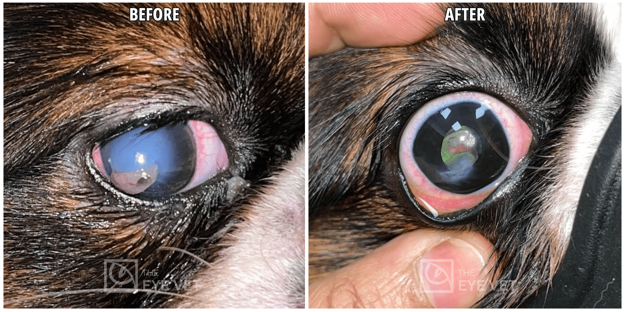
From emergency corneal tear suturing to advanced grafting and Gunderson’s flap surgery, we offer a spectrum of corneal surgeries designed to restore and protect your pet’s vision.

Corneal tear suturing
A penetrating injury to the cornea is a surgical emergency. If brought to us in time, we examine the eye completely and after thoroughly cleaning the eye, we resuture the cornea under an operating microscope.

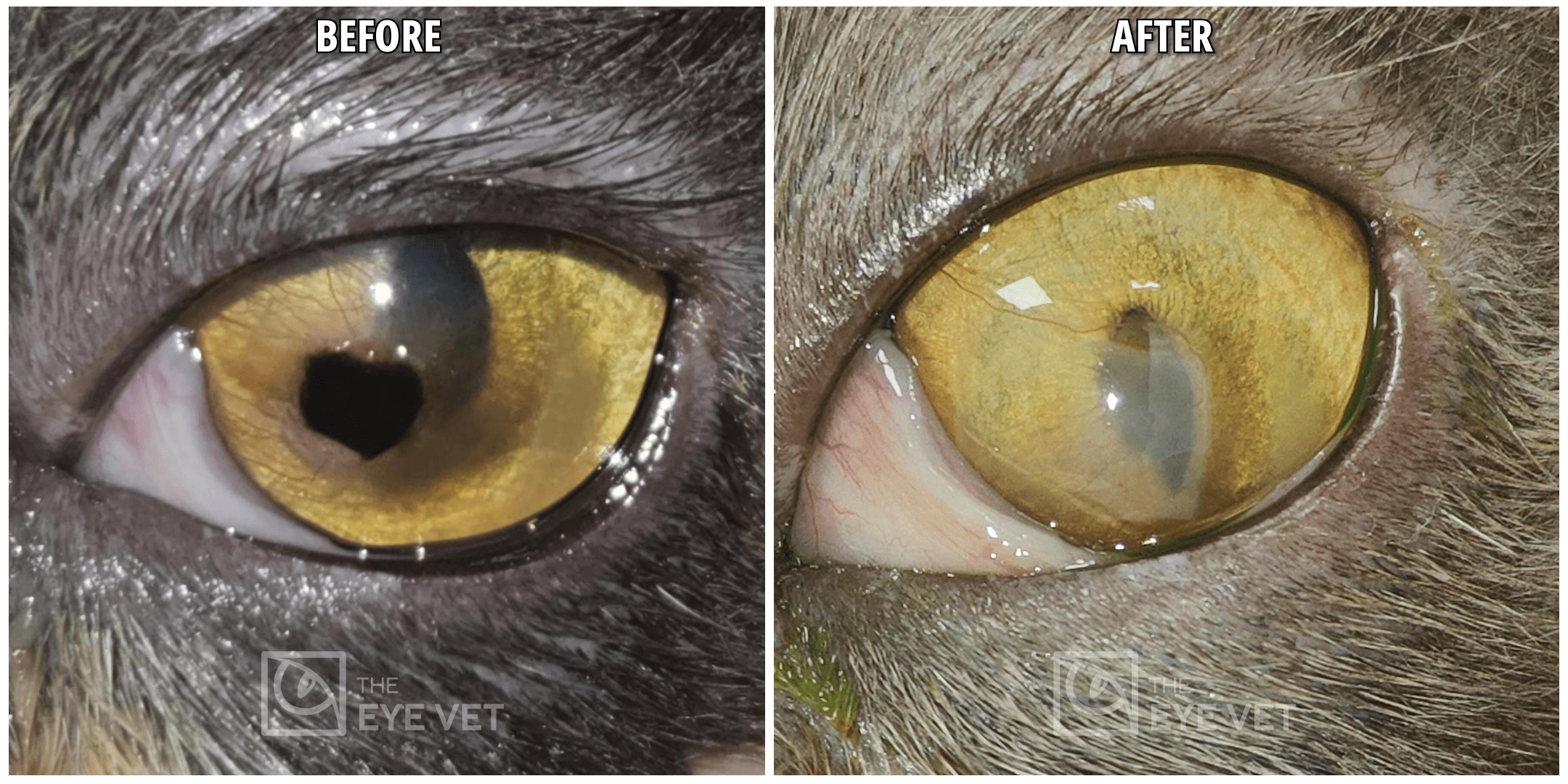
Corneal sequestrum removal in cats
Corneal sequestrum is a unique corneal degenerative condition. It appears like a dark brown pigment on the surface of the cornea. This is basically necrosis of the part of the cornea in response to a nonhealing corneal ulcer. Corneal sequestrum is a painful and progressive condition. Corneal sequestrum can go deeper in the cornea and can lead to perforation in effect leaving the eye blind. In some cats, the progression of sequestrum is very slow, whereas in some cats it can be rapid. Corneal sequestrum is treated surgically by removing the top layer of the cornea (keratectomy) involving the sequestrum. If the corneal defect is too deep after the keratectomy, we place a corneal graft in order to help heal the defect. If even a microscopic part of the necrosis is left behind, the sequestrum can regrow at the same spot.

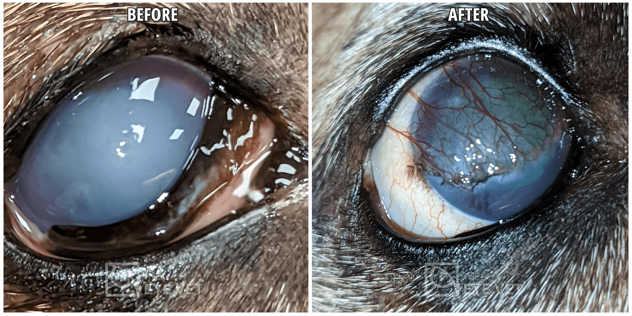
Gunderson Flap Technique
Corneal endothelial degeneration is a condition concerning the cornea's innermost cellular layer. Corneal endothelium is responsible for keeping the cornea clear by not allowing excess fluid to enter the cornea. With a finite number of endothelial cells present from birth, their loss can permit fluid infiltration into the cornea, resulting in corneal cloudiness. This may lead to non-healing corneal ulcers or even profound visual impairment as the cornea adopts an opaque, whitish hue. The Gunderson flap procedure surgically positions a conjunctival graft on the cornea. This procedure helps heal painful corneal ulcers and improves clarity of the cornea.

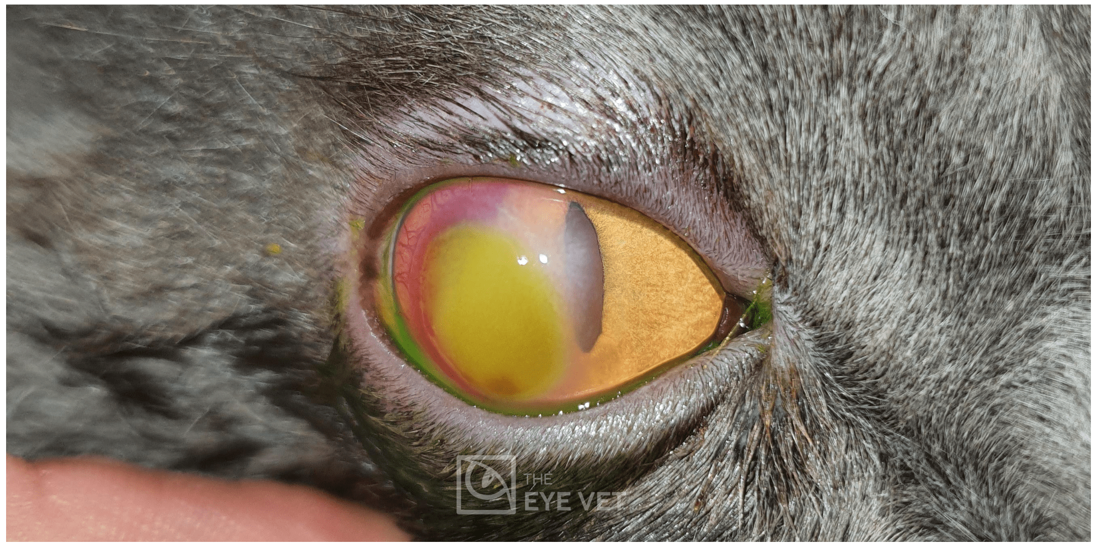
Corneal ulcers
A minor corneal wound heals within 3-5 days without much treatment. If the ulcer doesn’t heal within this time, it needs more aggressive treatment like:
Algerbrush debridement: A small round burr is used to roughen the surface of the ulcer. This is performed only in superficial corneal ulcers and a bandage contact lens is placed for the comfort of the patient. This procedure is done under topical anesthesia but if the patient is getting agitated, then light sedation can be used. Usually, with this treatment, 90% of the superficial ulcers heal. If the ulcer fails to heal with three consecutive algerbrush treatments, keratectomy surgery is suggested.
Keratectomy: is a surgical procedure performed under general anesthesia under an operating microscope. In this surgery, the surface layer of the corneal ulcer is removed and a bandage contact lens is placed. Success rate of this surgery is as high as 99%. Corneal Grafts: There are different types of corneal grafting procedures to help heal the deep ulcers:
a) Conjunctival pedicle graft: Thin conjunctiva over the white part of the eye is used to cover the ulcer and a thin pedicle is left attached to the white part which brings the blood and nourishment to the ulcer. This type of graft has a high success rate but this cannot be used in case of a ruptured ulcer. Disadvantage is that it leaves a significant scar at the site of surgery.
b) Corneo-conjunctival sliding graft: cornea adjacent to the ulcer is gently incised and slid over the ulcer. This graft is attached to the conjunctiva which brings in nutrition for the graft. The advantage of this graft over conjunctival graft is that the grafted portion after healing remains fairly transparent compared to conjunctival graft. It is a little more time consuming and if the cornea around the ulcer is not healthy, we have to use the other grafting materials. We prefer this technique for all the deep corneal ulcers.
c) Amnion graft: use of human amnion as a graft is a very successful procedure and leaves much less of a scar tissue compared to conjunctival graft. This graft cannot be used in deep or ruptured corneal ulcers.
d) Vetrix Biosis graft: This is a readymade graft made from porcine small intestine mucosa. We have imported this graft from Germany and our experience with this graft is very good. We use it to cover large deep corneal defects where extra tectonic support is important.
e) Full thickness cornea from other dogs: this is a good option for patients with a huge penetrating ulcer. The full thickness cornea can certainly fill the gap, but it hardly stays transparent (like in people). So this procedure is saved for instances when saving the globe is more important.
A penetrating injury to the cornea is a surgical emergency. If brought to us in time, we examine the eye completely and after thoroughly cleaning the eye, we resuture the cornea under an operating microscope.
A minor corneal wound heals within 3-5 days without much treatment. If the ulcer doesn’t heal within this time, it needs more aggressive treatment like:
Algerbrush debridement: A small round burr is used to roughen the surface of the ulcer. This is performed only in superficial corneal ulcers and a bandage contact lens is placed for the comfort of the patient. This procedure is done under topical anesthesia but if the patient is getting agitated, then light sedation can be used. Usually, with this treatment, 90% of the superficial ulcers heal. If the ulcer fails to heal with three consecutive algerbrush treatments, keratectomy surgery is suggested.
Keratectomy: is a surgical procedure performed under general anesthesia under an operating microscope. In this surgery, the surface layer of the corneal ulcer is removed and a bandage contact lens is placed. Success rate of this surgery is as high as 99%. Corneal Grafts: There are different types of corneal grafting procedures to help heal the deep ulcers:
a) Conjunctival pedicle graft: Thin conjunctiva over the white part of the eye is used to cover the ulcer and a thin pedicle is left attached to the white part which brings the blood and nourishment to the ulcer. This type of graft has a high success rate but this cannot be used in case of a ruptured ulcer. Disadvantage is that it leaves a significant scar at the site of surgery.
b) Corneo-conjunctival sliding graft: cornea adjacent to the ulcer is gently incised and slid over the ulcer. This graft is attached to the conjunctiva which brings in nutrition for the graft. The advantage of this graft over conjunctival graft is that the grafted portion after healing remains fairly transparent compared to conjunctival graft. It is a little more time consuming and if the cornea around the ulcer is not healthy, we have to use the other grafting materials. We prefer this technique for all the deep corneal ulcers.
c) Amnion graft: use of human amnion as a graft is a very successful procedure and leaves much less of a scar tissue compared to conjunctival graft. This graft cannot be used in deep or ruptured corneal ulcers.
d) Vetrix Biosis graft: This is a readymade graft made from porcine small intestine mucosa. We have imported this graft from Germany and our experience with this graft is very good. We use it to cover large deep corneal defects where extra tectonic support is important.
e) Full thickness cornea from other dogs: this is a good option for patients with a huge penetrating ulcer. The full thickness cornea can certainly fill the gap, but it hardly stays transparent (like in people). So this procedure is saved for instances when saving the globe is more important.
Corneal sequestrum is a unique corneal degenerative condition. It appears like a dark brown pigment on the surface of the cornea. This is basically necrosis of the part of the cornea in response to a nonhealing corneal ulcer. Corneal sequestrum is a painful and progressive condition. Corneal sequestrum can go deeper in the cornea and can lead to perforation in effect leaving the eye blind. In some cats, the progression of sequestrum is very slow, whereas in some cats it can be rapid. Corneal sequestrum is treated surgically by removing the top layer of the cornea (keratectomy) involving the sequestrum. If the corneal defect is too deep after the keratectomy, we place a corneal graft in order to help heal the defect. If even a microscopic part of the necrosis is left behind, the sequestrum can regrow at the same spot.
Endothelium is the innermost layer of the cornea. Animals are born with a fixed number of endothelial cells. If these cells start degenerating, fluid starts entering the cornea which turns it cloudy. In some cases the fluid retention in the cornea causes severe non healing corneal ulcers and in some cases the cornea becomes so white that it leads to severe visual deficits. In this surgery, we place a thin conjunctival graft on the surface of the cornea which drains the fluid from the eye and makes it transparent enough to improve vision in the affected animals.
